Description: Homo sapiens ATPase, Cu++ transporting, beta polypeptide (ATP7B), transcript variant 3, mRNA. RefSeq Summary (NM_001243182): This gene is a member of the P-type cation transport ATPase family and encodes a protein with several membrane-spanning domains, an ATPase consensus sequence, a hinge domain, a phosphorylation site, and at least 2 putative copper-binding sites. This protein is a monomer, and functions as a copper-transporting ATPase which exports copper out of the cells, such as the efflux of hepatic copper into the bile. Alternate transcriptional splice variants, encoding different isoforms with distinct cellular localizations, have been characterized. Mutations in this gene have been associated with Wilson disease which is characterized by copper accumulation. [provided by RefSeq, Dec 2019]. Transcript (Including UTRs) Position: hg19 chr13:52,506,806-52,534,146 Size: 27,341 Total Exon Count: 14 Strand: - Coding Region Position: hg19 chr13:52,508,892-52,532,617 Size: 23,726 Coding Exon Count: 14
ID:ATP7B_HUMAN DESCRIPTION: RecName: Full=Copper-transporting ATPase 2; EC=3.6.3.4; AltName: Full=Copper pump 2; AltName: Full=Wilson disease-associated protein; Contains: RecName: Full=WND/140 kDa; FUNCTION: Involved in the export of copper out of the cells, such as the efflux of hepatic copper into the bile. CATALYTIC ACTIVITY: ATP + H(2)O + Cu(2+)(In) = ADP + phosphate + Cu(2+)(Out). SUBUNIT: Monomer. Interacts with COMMD1/MURR1. Interacts with DCTN4, in a copper-dependent manner. Interacts with ATOX1. SUBCELLULAR LOCATION: Golgi apparatus, trans-Golgi network membrane; Multi-pass membrane protein (By similarity). Note=Predominantly found in the trans-Golgi network (TGN). Not redistributed to the plasma membrane in response to elevated copper levels. SUBCELLULAR LOCATION: Isoform 2: Cytoplasm. SUBCELLULAR LOCATION: WND/140 kDa: Mitochondrion. TISSUE SPECIFICITY: Most abundant in liver and kidney and also found in brain. Isoform 2 is expressed in brain but not in liver. The cleaved form WND/140 kDa is found in liver cell lines and other tissues. DOMAIN: Each HMA domain can bind a copper ion, they are tightly packed and closely interact with each other. Wild-type ATP7B can usually be loaded with an average 5.5 copper atoms per molecule. PTM: Isoform 1 may be proteolytically cleaved at the N-terminus to produce the WND/140 kDa form. DISEASE: Defects in ATP7B are the cause of Wilson disease (WD) [MIM:277900]. WD is an autosomal recessive disorder of copper metabolism in which copper cannot be incorporated into ceruloplasmin in liver, and cannot be excreted from the liver into the bile. Copper accumulates in the liver and subsequently in the brain and kidney. The disease is characterized by neurologic manifestations and signs of cirrhosis. SIMILARITY: Belongs to the cation transport ATPase (P-type) (TC 3.A.3) family. Type IB subfamily. SIMILARITY: Contains 6 HMA domains. SEQUENCE CAUTION: Sequence=AAA16173.1; Type=Frameshift; Positions=830; Sequence=AAA79211.1; Type=Frameshift; Positions=456; Sequence=AAA79212.1; Type=Frameshift; Positions=456; WEB RESOURCE: Name=GeneReviews; URL="http://www.ncbi.nlm.nih.gov/sites/GeneTests/lab/gene/ATP7B"; WEB RESOURCE: Name=Wilson Disease Mutation Database; URL="http://www.medicalgenetics.med.ualberta.ca/wilson/index.php";
arylsulfatase A pseudodeficiency Battisti C et al. 1999, Detection of a rare Wilson disease mutation associated with arylsulfatase A pseudodeficiency., American journal of medical genetics. 1999 Jul;85(2):175-8.
[PubMed 10406672]
liver disease; Wilson disease Liu, X. Q. et al. 2004, Correlation of ATP7B genotype with phenotype in Chinese patients with Wilson disease, World journal of gastroenterology. 2004 Feb;10(4):590-3.
[PubMed 14966923]
1384del17bp is a novel mutation found in WD patients. R778L is the most common mutation of ATP7B gene. There is a correlation between R778L and hepatic manifestations in WD patient.
Wilson disease Stapelbroek, J. M. et al. 2004, The H1069Q mutation in ATP7B is associated with late and neurologic presentation in Wilson disease:results of a meta-analysis., Journal of hepatology. 2004 Nov;41(5):758-63.
[PubMed 15519648]
Our results indicate that the H1069Q mutation is associated with a late and neurologic presentation.
The RNAfold program from the Vienna RNA Package is used to perform the secondary structure predictions and folding calculations. The estimated folding energy is in kcal/mol. The more negative the energy, the more secondary structure the RNA is likely to have.
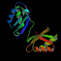
ModBase Predicted Comparative 3D Structure on P35670
Front
Top
Side
The pictures above may be empty if there is no ModBase structure for the protein. The ModBase structure frequently covers just a fragment of the protein. You may be asked to log onto ModBase the first time you click on the pictures. It is simplest after logging in to just click on the picture again to get to the specific info on that model.
Orthologous Genes in Other Species
Orthologies between human, mouse, and rat are computed by taking the best BLASTP hit, and filtering out non-syntenic hits. For more distant species reciprocal-best BLASTP hits are used. Note that the absence of an ortholog in the table below may reflect incomplete annotations in the other species rather than a true absence of the orthologous gene.