Human Gene SIGLEC10 (ENST00000353836.9) from GENCODE V44
Description: Homo sapiens sialic acid binding Ig like lectin 10 (SIGLEC10), transcript variant 3, mRNA. (from RefSeq NM_001171157) RefSeq Summary (NM_001171157): SIGLECs are members of the immunoglobulin superfamily that are expressed on the cell surface. Most SIGLECs have 1 or more cytoplasmic immune receptor tyrosine-based inhibitory motifs, or ITIMs. SIGLECs are typically expressed on cells of the innate immune system, with the exception of the B-cell expressed SIGLEC6 (MIM 604405).[supplied by OMIM, Jul 2002]. Gencode Transcript: ENST00000353836.9 Gencode Gene: ENSG00000142512.15 Transcript (Including UTRs) Position: hg38 chr19:51,410,021-51,417,803 Size: 7,783 Total Exon Count: 10 Strand: - Coding Region Position: hg38 chr19:51,411,099-51,417,581 Size: 6,483 Coding Exon Count: 10
ID:SIG10_HUMAN DESCRIPTION: RecName: Full=Sialic acid-binding Ig-like lectin 10; Short=Siglec-10; AltName: Full=Siglec-like protein 2; Flags: Precursor; FUNCTION: Putative adhesion molecule that mediates sialic-acid dependent binding to cells. Preferentially binds to alpha-2,3- or alpha-2,6-linked sialic acid. The sialic acid recognition site may be masked by cis interactions with sialic acids on the same cell surface. In the immune response, may act as an inhibitory receptor upon ligand induced tyrosine phosphorylation by recruiting cytoplasmic phosphatase(s) via their SH2 domain(s) that block signal transduction through dephosphorylation of signaling molecules. SUBUNIT: Interacts with PTPN6/SHP-1 upon phosphorylation. SUBCELLULAR LOCATION: Isoform 1: Cell membrane; Single-pass type I membrane protein. SUBCELLULAR LOCATION: Isoform 2: Cell membrane; Single-pass type I membrane protein. SUBCELLULAR LOCATION: Isoform 3: Cell membrane; Single-pass type I membrane protein. SUBCELLULAR LOCATION: Isoform 4: Cell membrane; Single-pass type I membrane protein. SUBCELLULAR LOCATION: Isoform 5: Secreted. TISSUE SPECIFICITY: Expressed by peripheral blood leukocytes (eosinophils, monocytes and a natural killer cell subpopulation). Isoform 5 is found to be the most abundant isoform. Found in lymph node, lung, ovary and appendix. Isoform 1 is found at high levels and isoform 2 at lower levels in bone marrow, spleen and spinal chord. Isoform 2 is also found in brain. Isoform 4 is specifically found in natural killer cells. DOMAIN: Contains 1 copy of a cytoplasmic motif that is referred to as the immunoreceptor tyrosine-based inhibitor motif (ITIM). This motif is involved in modulation of cellular responses. The phosphorylated ITIM motif can bind the SH2 domain of several SH2- containing phosphatases. PTM: Phosphorylation of Tyr-667 is involved in binding to PTPN6. SIMILARITY: Belongs to the immunoglobulin superfamily. SIGLEC (sialic acid binding Ig-like lectin) family. SIMILARITY: Contains 3 Ig-like C2-type (immunoglobulin-like) domains. SIMILARITY: Contains 1 Ig-like V-type (immunoglobulin-like) domain. WEB RESOURCE: Name=Functional Glycomics Gateway - Glycan Binding; Note=Siglec-10 [5 Fc Domains]; URL="http://www.functionalglycomics.org/glycomics/GBPServlet?&operationType=view&cbpId=cbp_hum_Itlect_00002"; WEB RESOURCE: Name=Functional Glycomics Gateway - Glycan Binding; Note=Siglec-10 long; URL="http://www.functionalglycomics.org/glycomics/GBPServlet?&operationType=view&cbpId=cbp_hum_Itlect_268";
The RNAfold program from the Vienna RNA Package is used to perform the secondary structure predictions and folding calculations. The estimated folding energy is in kcal/mol. The more negative the energy, the more secondary structure the RNA is likely to have.
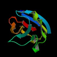
ModBase Predicted Comparative 3D Structure on Q96LC7
Front
Top
Side
The pictures above may be empty if there is no ModBase structure for the protein. The ModBase structure frequently covers just a fragment of the protein. You may be asked to log onto ModBase the first time you click on the pictures. It is simplest after logging in to just click on the picture again to get to the specific info on that model.
Orthologous Genes in Other Species
Orthologies between human, mouse, and rat are computed by taking the best BLASTP hit, and filtering out non-syntenic hits. For more distant species reciprocal-best BLASTP hits are used. Note that the absence of an ortholog in the table below may reflect incomplete annotations in the other species rather than a true absence of the orthologous gene.