Human Gene CABP1 (ENST00000316803.8) from GENCODE V44
Description: Homo sapiens calcium binding protein 1 (CABP1), transcript variant 3, mRNA. (from RefSeq NM_001033677) RefSeq Summary (NM_001033677): Calcium binding proteins are an important component of calcium mediated cellular signal transduction. This gene encodes a protein that belongs to a subfamily of calcium binding proteins which share similarity to calmodulin. The protein encoded by this gene regulates the gating of voltage-gated calcium ion channels. This protein inhibits calcium-dependent inactivation and supports calcium-dependent facilitation of ion channels containing voltage-dependent L-type calcium channel subunit alpha-1C. This protein also regulates calcium-dependent activity of inositol 1,4,5-triphosphate receptors, P/Q-type voltage-gated calcium channels, and transient receptor potential channel TRPC5. This gene is predominantly expressed in retina and brain. Alternative splicing results in multiple transcript variants encoding disinct isoforms. [provided by RefSeq, Jul 2012]. Gencode Transcript: ENST00000316803.8 Gencode Gene: ENSG00000157782.10 Transcript (Including UTRs) Position: hg38 chr12:120,640,626-120,667,324 Size: 26,699 Total Exon Count: 6 Strand: + Coding Region Position: hg38 chr12:120,640,686-120,666,900 Size: 26,215 Coding Exon Count: 6
ID:CABP1_HUMAN DESCRIPTION: RecName: Full=Calcium-binding protein 1; Short=CaBP1; AltName: Full=Calbrain; AltName: Full=Caldendrin; FUNCTION: Modulates calcium-dependent activity of inositol 1,4,5- triphosphate receptors (ITPRs). Inhibits agonist-induced intracellular calcium signaling. Enhances inactivation and does not support calcium-dependent facilitation of voltage-dependent P/Q-type calcium channels. Causes calcium-dependent facilitation and inhibits inactivation of L-type calcium channels by binding to the same sites as calmodulin in the C-terminal domain of CACNA1C, but resulting in an opposit effects on channel function. Suppresses the calcium-dependent inactivation of CACNA1D (By similarity). Inhibits TRPC5 channels. Prevents NMDA receptor- induced cellular degeneration (By similarity). SUBUNIT: Homodimer; when bound to calcium or magnesium. Interacts (via C-terminus) with ITPR1, ITPR2 and ITPR3. This binding is calcium dependent and the interaction correlates with calcium concentration. An additional calcium-independent interaction with the N-terminus of ITPR1 results in a decreased InsP(3) binding to the receptor. Interacts with CACNA1A (via C-terminal CDB motif) in the pre- and postsynaptic membranes. Interacts with CACNA1C (via C-terminal C and IQ motifs). The binding to the C motif is calcium independent whereas the binding to IQ requires the presence of calcium and is mutually exclusive with calmodulin binding. Interacts with CACNA1D (By similarity). Interacts with TRPC5 (via C-terminus). Interacts (via EF-hands 1 and 2) at microtubules with MAP1LC3B. Interacts with MYO1C and NELF (By similarity). INTERACTION: Q13936:CACNA1C; NbExp=4; IntAct=EBI-907894, EBI-1038838; SUBCELLULAR LOCATION: Cytoplasm, cytoskeleton. Cytoplasm, perinuclear region. Cell membrane; Lipid-anchor; Cytoplasmic side. Golgi apparatus. Cell junction, synapse, postsynaptic cell membrane, postsynaptic density. Note=L-CaBP1 is associated most likely with the cytoskeletal structures, whereas S-CaBP1 is localized at or near the plasma membrane. SUBCELLULAR LOCATION: Isoform L-CaBP1: Cytoplasm, cytoskeleton. Note=L-CaBP1 is associated most likely with the cytoskeletal structures. SUBCELLULAR LOCATION: Isoform S-CaBP1: Cytoplasm, cell cortex. Cell membrane; Lipid-anchor (Probable). Note=S-CaBP1 is localized at or near the plasma membrane. TISSUE SPECIFICITY: Retina and brain. Somatodendritic compartment of neurons. Calbrain was found exclusively in brain where it is abundant in the hippocampus, habenular area in the epithalamus and in the cerebellum. DOMAIN: EF-1 binds magnesium constitutively under physiological conditions, EF-3 and EF-4 bind calcium cooperatively and EF-2 binds neither calcium nor magnesium. PTM: Phosphorylated. The phosphorylation regulates the activity. SIMILARITY: Contains 4 EF-hand domains. CAUTION: The interaction with CACNA1A is described as calcium independent in PubMed:11865310 while it is shown to be acutely calcium dependent in PubMed:14570872. PubMed:12032348 describes a stimulatory effect of CABP1 during agonist-induced intracellular calcium signaling while PubMed:14570872 and PubMed:11865310 show an inhibitory effect.
The RNAfold program from the Vienna RNA Package is used to perform the secondary structure predictions and folding calculations. The estimated folding energy is in kcal/mol. The more negative the energy, the more secondary structure the RNA is likely to have.
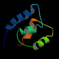
ModBase Predicted Comparative 3D Structure on Q9NZU7
Front
Top
Side
The pictures above may be empty if there is no ModBase structure for the protein. The ModBase structure frequently covers just a fragment of the protein. You may be asked to log onto ModBase the first time you click on the pictures. It is simplest after logging in to just click on the picture again to get to the specific info on that model.
Orthologous Genes in Other Species
Orthologies between human, mouse, and rat are computed by taking the best BLASTP hit, and filtering out non-syntenic hits. For more distant species reciprocal-best BLASTP hits are used. Note that the absence of an ortholog in the table below may reflect incomplete annotations in the other species rather than a true absence of the orthologous gene.
Biological Process: GO:0007601 visual perception GO:0042308 negative regulation of protein import into nucleus GO:0043086 negative regulation of catalytic activity