ID:JAM3_HUMAN DESCRIPTION: RecName: Full=Junctional adhesion molecule C; Short=JAM-C; AltName: Full=JAM-2; AltName: Full=Junctional adhesion molecule 3; Short=JAM-3; Flags: Precursor; FUNCTION: Participates in cell-cell adhesion. It is a counter- receptor for ITGAM, mediating leukocyte-platelet interactions and is involved in the regulation of transepithelial migration of polymorphonuclear neutrophils (PMN). The soluble form is a mediator of angiogenesis. SUBUNIT: Interacts with JAM2. Interacts with ITGAM. INTERACTION: P57087:JAM2; NbExp=2; IntAct=EBI-4314733, EBI-3918416; SUBCELLULAR LOCATION: Cell membrane; Single-pass type I membrane protein (Potential). Cell junction, desmosome. Secreted, extracellular space. Note=In epithelial cells, it is expressed at desmosomes but not at tight junctions. Localizes at the cell surface of endothelial cells; treatment of endothelial cells with vascular endothelial growth factor stimulates recruitment of JAM3 to cell-cell contacts. TISSUE SPECIFICITY: Highest expression in placenta, brain and kidney. Significant expression is detected on platelets. Expressed in intestinal mucosa cells. Expressed in the vascular endothelium. Found in serum (at protein level). Also detected in the synovial fluid of patients with rheumatoid arthritis, psoriatic arthritis or ostearthritis (at protein level). PTM: Proteolytically cleaved from endothelial cells surface into a soluble form by ADAM10 and ADAM17; the release of soluble JAM3 is increased by proinflammatory factors. DISEASE: Defects in JAM3 are the cause of hemorrhagic destruction of the brain with subependymal calcification and cataracts (HDBSCC) [MIM:613730]. A syndrome characterized by congenital cataracts and severe brain abnormalities apparently resulting from hemorrhagic destruction of the brain tissue, including the cerebral white matter and basal ganglia. Patients manifest profound developmental delay, and other neurologic features included seizures, spasticity, and hyperreflexia. Brain imaging shows multifocal intraparenchymal hemorrhage with associated liquefaction and massive cystic degeneration, and calcification in the subependymal region and in brain tissue. SIMILARITY: Belongs to the immunoglobulin superfamily. SIMILARITY: Contains 1 Ig-like C2-type (immunoglobulin-like) domain. SIMILARITY: Contains 1 Ig-like V-type (immunoglobulin-like) domain. SEQUENCE CAUTION: Sequence=CAC94776.1; Type=Erroneous initiation;
The RNAfold program from the Vienna RNA Package is used to perform the secondary structure predictions and folding calculations. The estimated folding energy is in kcal/mol. The more negative the energy, the more secondary structure the RNA is likely to have.
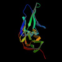
ModBase Predicted Comparative 3D Structure on Q9BX67
Front
Top
Side
The pictures above may be empty if there is no ModBase structure for the protein. The ModBase structure frequently covers just a fragment of the protein. You may be asked to log onto ModBase the first time you click on the pictures. It is simplest after logging in to just click on the picture again to get to the specific info on that model.
Orthologous Genes in Other Species
Orthologies between human, mouse, and rat are computed by taking the best BLASTP hit, and filtering out non-syntenic hits. For more distant species reciprocal-best BLASTP hits are used. Note that the absence of an ortholog in the table below may reflect incomplete annotations in the other species rather than a true absence of the orthologous gene.
 Sequence and Links to Tools and Databases

 Microarray Expression Data