Human Gene CLDN7 (ENST00000360325.11) from GENCODE V44
Description: Homo sapiens claudin 7 (CLDN7), transcript variant 1, mRNA. (from RefSeq NM_001307) RefSeq Summary (NM_001307): This gene encodes a member of the claudin family. Claudins are integral membrane proteins and components of tight junction strands. Tight junction strands serve as a physical barrier to prevent solutes and water from passing freely through the paracellular space between epithelial or endothelial cell sheets, and also play critical roles in maintaining cell polarity and signal transductions. Differential expression of this gene has been observed in different types of malignancies, including breast cancer, ovarian cancer, hepatocellular carcinomas, urinary tumors, prostate cancer, lung cancer, head and neck cancers, thyroid carcinomas, etc.. Alternatively spliced transcript variants encoding different isoforms have been found.[provided by RefSeq, May 2010]. Gencode Transcript: ENST00000360325.11 Gencode Gene: ENSG00000181885.18 Transcript (Including UTRs) Position: hg38 chr17:7,259,903-7,262,478 Size: 2,576 Total Exon Count: 4 Strand: - Coding Region Position: hg38 chr17:7,260,374-7,262,043 Size: 1,670 Coding Exon Count: 4
ID:CLD7_HUMAN DESCRIPTION: RecName: Full=Claudin-7; Short=CLDN-7; FUNCTION: Plays a major role in tight junction-specific obliteration of the intercellular space (By similarity). SUBUNIT: Directly interacts with TJP1/ZO-1, TJP2/ZO-2 and TJP3/ZO- 3 (By similarity). The phosphorylated form interacts with EPCAM. SUBCELLULAR LOCATION: Cell membrane; Multi-pass membrane protein. Lateral cell membrane. Cell junction, tight junction. Note=Co- localizes with EPCAM at the lateral cell membrane and tight junction. TISSUE SPECIFICITY: Expressed in kidney, lung and prostate. Isoform 1 seems to be predominant, except in some normal prostate samples, where isoform 2 is the major form. Down-regulated in breast cancers, including ductal carcinoma in situ (DCIS), lobular carcinoma in situ (LCIS) and invasive ductal carcinoma (IDC) (at protein level), as well as in several cancer cell lines. Loss of expression correlates with histological grade, occurring predominantly in high-grade lesions. INDUCTION: By androgens. PTM: Phosphorylated. SIMILARITY: Belongs to the claudin family. SEQUENCE CAUTION: Sequence=AAP97219.1; Type=Frameshift; Positions=18, 23;
The RNAfold program from the Vienna RNA Package is used to perform the secondary structure predictions and folding calculations. The estimated folding energy is in kcal/mol. The more negative the energy, the more secondary structure the RNA is likely to have.
Pfam Domains: PF00822 - PMP-22/EMP/MP20/Claudin family
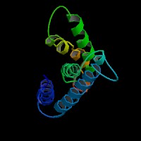
ModBase Predicted Comparative 3D Structure on O95471
Front
Top
Side
The pictures above may be empty if there is no ModBase structure for the protein. The ModBase structure frequently covers just a fragment of the protein. You may be asked to log onto ModBase the first time you click on the pictures. It is simplest after logging in to just click on the picture again to get to the specific info on that model.
Orthologous Genes in Other Species
Orthologies between human, mouse, and rat are computed by taking the best BLASTP hit, and filtering out non-syntenic hits. For more distant species reciprocal-best BLASTP hits are used. Note that the absence of an ortholog in the table below may reflect incomplete annotations in the other species rather than a true absence of the orthologous gene.
Gene Ontology (GO) Annotations with Structured Vocabulary
Molecular Function: GO:0005198 structural molecule activity GO:0005515 protein binding GO:0042802 identical protein binding GO:0050839 cell adhesion molecule binding
Biological Process: GO:0007162 negative regulation of cell adhesion GO:0008284 positive regulation of cell proliferation GO:0016338 calcium-independent cell-cell adhesion via plasma membrane cell-adhesion molecules GO:0032463 negative regulation of protein homooligomerization GO:0043066 negative regulation of apoptotic process GO:0045471 response to ethanol GO:2000147 positive regulation of cell motility